Comparison: X-Ray vs Ultrasonic Testing for Castings
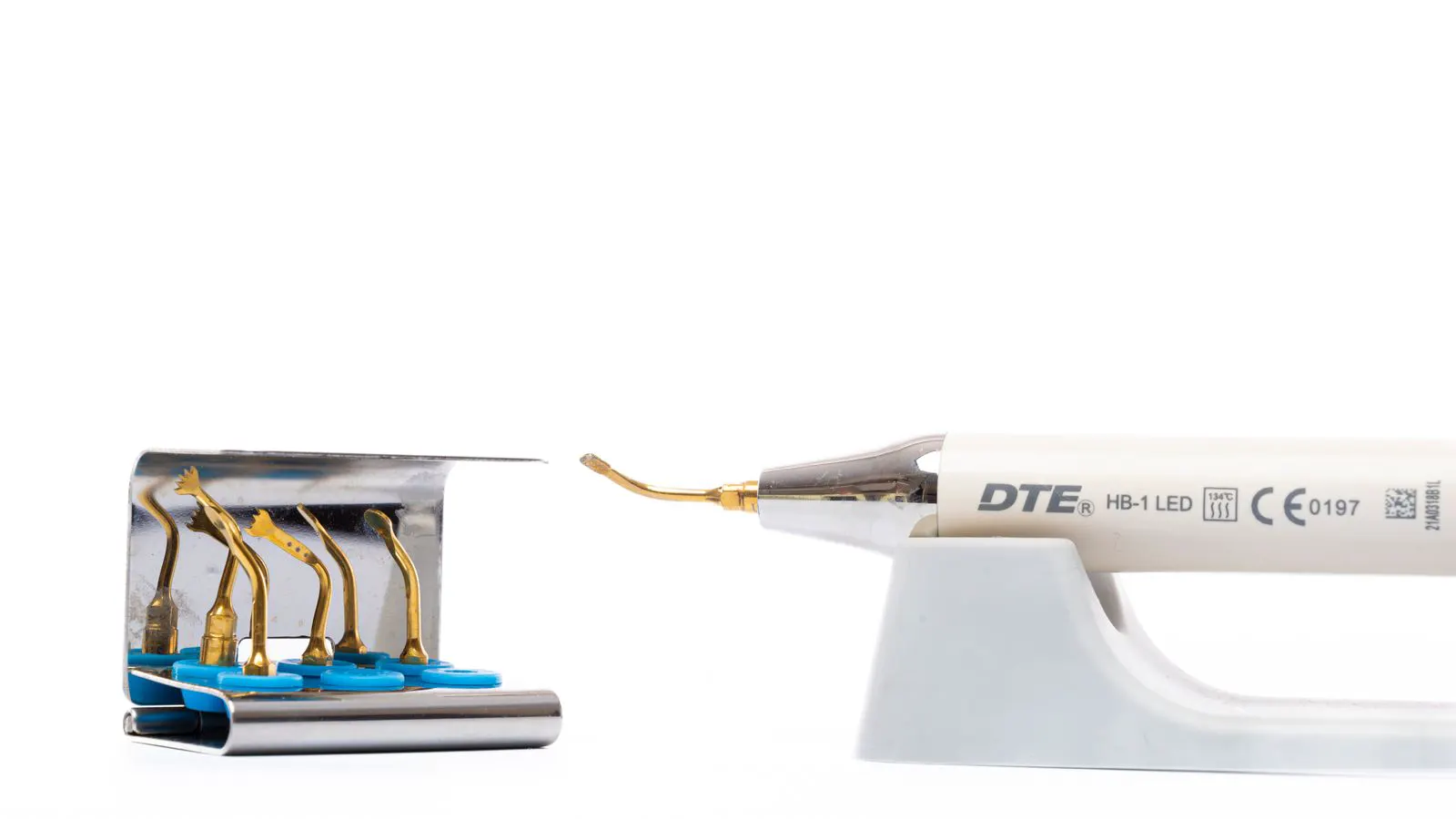
X-ray and ultrasonic testing are the two primary methods for finding internal defects in investment castings. X-ray excels with dense, thick sections, while ultrasonic is faster and cheaper for complex geometries. Select based on material, size, and defect type.
- X-ray provides a 2D image of internal voids and porosity but struggles with dense materials.
- Ultrasonic testing detects subsurface cracks and voids quickly but requires good coupling and access.
- Dense alloys and very thick sections favor X-ray, while complex shapes and high-volume parts favor ultrasonic.
- Combining both methods often gives the most complete picture of internal casting quality.
How to Choose Between X-Ray and Ultrasonic Testing for Investment Castings
Engineers selecting non-destructive testing methods must match the technique to the part, the material, and the defect risk. For investment castings, the choice between X-ray and ultrasonic testing often determines whether a hidden defect is caught before the part reaches assembly.
What Each Method Actually Detects
X-ray and ultrasonic testing do not measure the same thing in the same way. X-ray imaging creates a 2D projection of the entire part. It shows density variations. A hot tear, a large shrinkage pore, or a sand inclusion will appear as a darker or lighter area against the background density of the metal. The image is a slice through the part. If two defects overlap along the X-ray beam path, they merge into one shadow.
Ultrasonic testing works differently. A transducer sends high-frequency sound waves into the casting. The sound travels through the metal and reflects off boundaries, voids, or cracks. The receiver measures the time and amplitude of the echo. This method maps thickness and internal discontinuities along the beam path. It is more directional than X-ray. It can detect a crack that is parallel to the surface, or a void that is perpendicular to the sound wave, with high sensitivity.
The core difference is geometry. X-ray is an all-around view through the part. Ultrasonic is a line-by-line scan that must be aimed at the expected defect location.
Consider a gearbox housing made from A356 aluminum. The material has a relatively low acoustic impedance, meaning sound waves travel through it with less loss. An ultrasonic probe can easily detect a cluster of microporosity near the core lines. If the same part were made from 4130 chrome-moly steel, the acoustic signal would attenuate much faster. The same cluster of porosity might produce a weak, noisy echo. An X-ray image, however, would show the density contrast clearly because the steel absorbs X-rays predictably. The material dictates the physics of the signal, and the physics dictates the limit of detection.
X-Ray vs Ultrasonic: Comparison Table
The table below summarizes the core trade-offs between the two methods for internal defect detection.
| Option | Best for | Limitations |
|---|---|---|
| X-Ray | Dense materials, thick sections, mapping porosity | 2D projection, overlapping defects, high cost for large parts |
| Ultrasonic | Cracks, complex shapes, high-volume inspection | Requires coupling, access issues, operator skill dependent |
| Radiography | Parts with high density variations | Shadow overlap, cannot measure depth accurately |
| Emission Acoustic | Thin-walled sections, small parts | Limited penetration depth, surface sensitive |
X-ray is the go-to method when the material is dense and the section is thick. Lead, brass, and many steels attenuate sound waves significantly. In these cases, ultrasonic signals degrade quickly. X-rays penetrate deep into the material. The image reveals the internal structure without needing to touch the surface.
Ultrasonic testing shines when the part is complex. Investment castings often have intricate internal features. A transducer can be placed on an accessible flat surface and angled to scan a hidden cavity. X-ray would require the part to be mounted in a specific orientation to capture the critical area.
When to Pick X-Ray
Pick X-ray when the defect is a density anomaly. Shrinkage porosity is the most common internal defect in investment casting. It forms during solidification and leaves small gas pockets behind. These pockets have a different density than the surrounding metal. X-ray captures them well.
X-ray is also the better choice for very thick sections. If a casting has a section thickness of over two inches, ultrasonic waves may attenuate too much to give a reliable echo. The signal-to-noise ratio drops. The operator may miss small voids. X-ray does not have this penetration limit. It can see through thick steel without loss of image quality.
The cost is higher. X-ray requires a lead shield, a lead apron, and a controlled area. The part must be moved into the X-ray room. For a large production run, the throughput is slower. Each part takes time to position and expose.
Consider a turbine impeller made from Inconel 718. The alloy is dense and the wall thickness is substantial. An ultrasonic scan might show strong reflections from the surface, but the signal will fade before it reaches the root of the blade. The operator might see a clean profile while the root is full of micro-shrinkage. An X-ray film, however, captures the entire volume. The density contrast of the Inconel against the air in the pores is sharp. The image reveals the porosity pattern even in the deepest sections.
When to Pick Ultrasonic
Pick ultrasonic when the defect is a crack or a void. Cracks are linear discontinuities. They reflect sound waves strongly. A hot tear or a cold shut, which are common in investment casting, create a boundary that the ultrasonic beam detects. The echo is sharp and easy to identify.
Ultrasonic testing is faster. It can be done on the production floor. No X-ray room is required. A portable unit with a transducer and a coupling gel is all you need. This makes it ideal for high-volume inspection. You can scan a batch of parts in a short time.
The operator skill matters. Ultrasonic is not a “set it and forget it” method. The operator must angle the transducer correctly. A small change in angle can miss a crack that is parallel to the surface. The coupling gel must be applied evenly. Air gaps between the gel and the metal kill the signal.
Think of a hydraulic pump housing made from gray iron. The part has a large, flat bottom face. A crack formed during the gating process might run horizontally near the bottom. An X-ray image would show a faint shadow if the crack is thin enough to catch the beam. An ultrasonic probe, placed on the bottom face and angled at 45 degrees, sends waves directly into the crack plane. The reflection is a sharp spike on the display. The operator can measure the depth of the crack to the millimeter. The speed of this check allows the foundry to reject the bad part before machining begins.
Practical Limitations of Each Method
Both methods have limits. X-ray is a 2D projection. If a casting has a complex shape, the internal features overlap. A large void and a small crack might appear as one dark blob. You cannot tell how deep the defect is. You only know it is there.
Ultrasonic testing requires good surface access. Investment castings often have rough surfaces. Gating remnants, sprues, and rough castings can scatter the sound waves. The operator must grind the area to get a smooth surface. This adds time and labor.
Also, ultrasonic testing is sensitive to the material. Cast iron is a poor conductor of sound waves. The porosity in the iron absorbs the energy. The echoes are weak. X-ray handles cast iron much better. It shows the density variations clearly.
Consider a complex automotive manifold made from aluminum. The part has many intersecting passages and thin walls. X-ray is excellent for showing the overall porosity in the thick walls. However, the 2D nature of the image makes it difficult to distinguish between a small pore in the front wall and a pore in the back wall. They overlap. An ultrasonic scan can be performed on the external surface to map the thickness. It can confirm whether the wall is uniform. If a defect is found, the ultrasonic method provides a depth coordinate. The X-ray image provides a location coordinate. Together, they define the 3D position of the defect.
How to Combine Both Methods
Many foundries use both methods. The approach is simple. Use X-ray to map the porosity. Use ultrasonic to check for cracks. The X-ray image gives a general picture of the internal structure. The ultrasonic scan confirms the location of linear defects.
This combination is most useful for critical parts. If a casting fails, the cost is high. The engineer wants to know if there are any hidden cracks or voids. The dual method provides a more complete safety margin.
The process is straightforward. First, X-ray the part. Review the image. If the porosity looks acceptable, move to ultrasonic. Scan the critical areas. If the ultrasonic scan shows no echoes, the part is likely clean.
For a medical implant or an aerospace bracket, the tolerance for defects is near zero. An X-ray inspection might pass a part with a small cluster of porosity that is technically within the acceptance limits for general industrial use. However, for a high-stress application, that porosity could be a stress riser. The engineer might then call for an ultrasonic scan to rule out any associated hot tears or cracks. The X-ray tells you the porosity is there. The ultrasonic tells you the metal around it is continuous.
Common Mistakes in Method Selection
Engineers often pick the wrong method based on habit. They assume X-ray is always better. Or they assume ultrasonic is always cheaper. Both are wrong.
A common mistake is using ultrasonic on a dense alloy without checking the material properties. The operator expects a good signal. The signal is weak. The part fails later. The defect was there, but the method was wrong.
Another mistake is neglecting the surface condition. Investment castings often have a rough finish. The operator does not grind the surface. The gel does not bond. The signal is noisy. The defect is missed.
The key is to match the method to the part. Look at the material, the size, and the defect risk. Do not default to one method without thinking.
A final error is ignoring the geometry of the defect. Engineers often design parts with thin walls that are prone to hot tears. They then request X-ray only. The X-ray shows a faint shadow. It is within the visual acceptance criteria. The part is released. In service, the hot tear propagates under cyclic loading. The part fails. Had an ultrasonic scan been performed on the thin wall, the linear crack would have been detected with high sensitivity. The defect type, not just the size, should drive the testing method.
In summary, the choice is not about which method is superior. It is about which method is appropriate for the specific physical characteristics of the casting and the specific type of defect that is likely to occur. Engineers must look at the material density, the wall thickness, the surface finish, and the criticality of the part. X-ray provides a global view of density. Ultrasonic provides a detailed view of linear discontinuities. When the stakes are high, both are necessary. When the stakes are low, one may be sufficient. The decision must be made based on physics and risk, not on convenience or habit.
Frequently asked questions
Can X-ray detect cracks in castings?
X-ray can detect cracks if they are dense enough to create a shadow. However, it is not the best method for thin cracks. Ultrasonic is more sensitive to linear discontinuities.
Is ultrasonic testing safer than X-ray?
Yes. Ultrasonic testing uses sound waves. There is no radiation. It can be done on the production floor. X-ray requires a shielded area and safety protocols.
Which method is better for cast iron?
X-ray is generally better for cast iron. The porosity in the iron attenuates sound waves. Ultrasonic signals are weak in cast iron. X-ray shows the density variations clearly.
Can I use both methods on the same part?
Yes. Many foundries use both. X-ray maps the porosity, and ultrasonic checks for cracks. This provides a more complete picture of the internal quality.
How much does it cost to test a casting?
Costs depend on the size of the part, the method, and the volume. X-ray is generally more expensive per part. Ultrasonic is cheaper and faster.